New Renaissance Institute has developed a family of lensless technologies that can inexpensively implement a variety of high-performance microscopic imaging and microscopic optical tomography functions without lenses. These technologies adapt and repurpose many of NRI Lensless Light Field Imaging technologies. Implementations can be extremely inexpensive, easily replicated, and in the limit can be fabricated with (semiconducting ink) printing or deposition techniques similar to that used to manufacture OLED (Organic Light Emitting Diode) displays. Depending on the material employed, the NRI illumination and/or imaging elements can even be (ecologically-responsibly) disposable or recycled.
Surprisingly, with proper selection and engineering of components, is possible to use a common sensor/illumination hardware arrangements to implement one, more, or all of these low-cost small-profile microscopic electronic digital imaging and tomography capabilities:
- NRI Lensless electronic transmission microscopy (inexpensive magnification of > 100x, no focusing needed)
- NRI Lensless electronic reflection microscopy (can provide self-illumination, can focus at “zero” separation distance)
- NRI Lensless electronic fluorescent microscopy (can include selectable-wavelength fluorescent-marker stimulation)
- NRI Lensless electronic transmission optical tomography.
Light can be (sequentially or simultaneously) sensed and emitted from the same surface (permitting ‘self-illumination”), and the imaging surface and/or focus-surface can be planar or curved. The transmission microscopy imaging methods employing collimated light require no focusing, and other (light-field) microscopy imaging approaches implement image formation in software and can focus under software control in real time or at a later time operating on stored data. Using microLED-like, OLED-like, and other reciprocal optoelectronic elements as light sensors permit wavelength-dependent responses, facilitating the implementation of color and broader-range multi-band imaging without any need for optical filters or diffraction gradings. Use of OLED arrays and micro-LED arrays can be used to create wavelength-differentiating self-illuminating image sensors for reflective microscopy and sequential-scanning illumination arrays for use in the transmission optical tomography modalities.
To create images for microscopy or optical tomography, an imaging sensor needs an associated light source to illuminate the wells.
- In NRI lensless transmission microscopy, illumination is provided on the opposite side of a translucent specimen being imaged so that the illumination light from the light source travels through the translucent specimen rather than being reflected by the translucent specimen. Most light sources emit a geometrically diverging uncollimated light field as the light propagates from the light source. To capture sharp images in NRI’s lensless transmission microscopy approaches, the lensless imaging sensor employs a miniaturized collimated light source or approximating arrangements.
- In NRI lensless reflection microscopy, illumination is provided on the same side of the translucent specimen being imaged rather on the opposite side of the translucent specimen being imaged so that the illumination light from the light source is reflected off the translucent specimen. NRI lensless reflection microscopy approaches employ NRI’s Lensless Light Field Imaging approaches implemented for microscopic imaging.
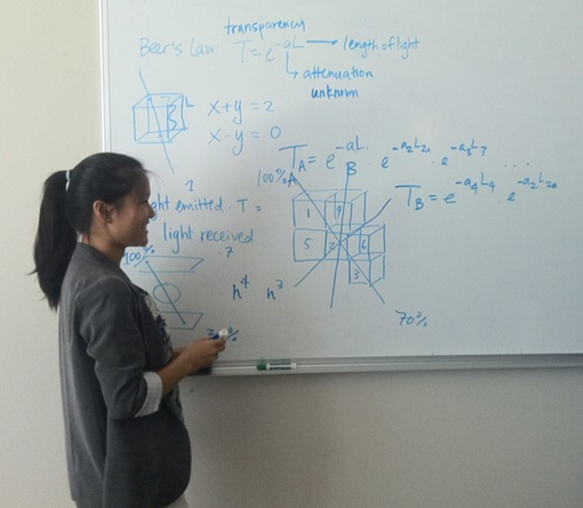
- NRI optical tomography creates a 3D lattice (voxel) representation of a translucent object computed from light transmitted and scattered through an object. Individual light-emitting elements of a sequentially-illuminated light source array emits a spreading light pattern in various locations within the image sensor. NRI optical tomography approaches represent the translucent object as a 3-dimensional array of voxels, each voxel having a homogeneous opacity for the wavelength of light emitted from light source. Light paths between light sources and image sensor pixels intersect different groups of voxels as the light travels through (and to varying degrees scatters and reflects within) the sample. As light propagates through the translucent sample, (Beer-Lambert) exponential light attention invoked by light absorption processes. By applying logarithmic operations to measured data sets across a sufficient number of light paths, an adequate number of linear equations result that can be solved with standard matrix techniques for the log-values of opacities of each voxel, from which the opacity values of each voxel can be obtained by exponentiating the linear equation solutions. Scattering and dark objects introduce distortion effects which can be addressed to various degrees by additional mathematical and computational treatment.
A broad range of such next-generation imaging capabilities can be miniaturized and manufactured so inexpensively it becomes possible to provided separate dedicated advanced multifunction microscopic imaging operating simultaneously for each well in an (ANSI standard) 24-well, 96-well, or even 384-well microplate providing magnifications of up to 110X or greater. Accordingly, NRI is currently directing this work to inexpensive simultaneous non-robotic imaging of every well in (living cell-culture) microplates for non-removal operation within a CO2 incubator (see Next-Generation Microplate Technologies); these adaptations are being licensed by Advanced Microplate Technologies.
Issued Patents
| Title | Patent Number | Application Number | Priority Dates | Text Only | Related Patents | |
|---|---|---|---|---|---|---|
| Optical Tomography Optoelectronic Arrangements For Microplate Wells | 10,816,467 | 16/036,646 | 08/09/2013 | Text | Proximate Microscopic Imaging and Tomography | |
| Optical Tomography Optoelectronic Arrangements for Microscopy, Cell Cytometry, Microplate Array Instrumentation, Crystallography, and Other Applications | 10,656,076 | 15/652,141 | 06/16/2009 06/16/2010 08/09/2013 | Text | Proximate Microscopic Imaging and Tomography | |
| Small-Profile Lensless Optical Microscopy Imaging and Tomography Instruments and Elements For Low Cost And Integrated Microscopy | 10,498,939 | 14/105,108 | 06/16/2009 06/16/2010 12/12/2013 | Text | Proximate Microscopic Imaging and Tomography | |
| Cylindrical Optical Tomography for Microscopy, Cell Cytometry, Microplate Array Instrumentation, Crystallography, and Other Applications | 10,024,791 | 15/457,963 | 08/09/2013 | Text | Proximate Microscopic Imaging and Tomography | |
| Electronic imaging flow-microscope for environmental remote sensing, bioreactor process monitoring, and optical microscopic tomography | 9,709,483 | 15/289,815 | 06/16/2009 | Text | Proximate Microscopic Imaging and Tomography |
|
| Optical tomography for microscopy, cell cytometry, microplate array instrumentation, crystallography, and other applications | 9,594,239 | 13/963,917 | 06/16/2009 06/16/2010 08/09/2013 | Text | Proximate Microscopic Imaging and Tomography |
|
| Optical tomography for microscopy, cell cytometry, microplate array instrumentation, crystallography, and other applications | 9,594,019 | 13/963,931 | 08/09/2013 | Text | Proximate Microscopic Imaging and Tomography |
|
| Electronic imaging flow-microscope for environmental remote sensing, bioreactor process monitoring, and optical microscopic tomography | 8,885,035 | 12/817,107 | 06/16/2009 | Text | Proximate Microscopic Imaging and Tomography |
Pending Published Applications
| Title | Publication Number | Application Number | Priority Dates | Publish Date | Text Only | Related Patents |
Pending Unpublished Applications
| Title | Application Number | Priority Dates | Related Patents |
|---|---|---|---|
| Optical Tomography Optoelectronic Arrangements for Advanced Microscopy Applications | 17/081,992 | 08/09/2013 | Proximate Microscopic Imaging and Tomography |
| Computational Optical Tomography Optoelectronic Arrangements for Microscopy, Cell Cytometry, Microplate Array Instrumentation, Crystallography, and Other Applications | 16/878,572 | 06/16/2009 | Proximate Microscopic Imaging and Tomography |
| Small-Profile Lensless Optical Tomography and Lensless Microscopic Imaging Arrangements for Low Cost Instrumentation and Integration | 16/700,798 | 06/16/09 12/12/13 | Proximate Microscopic Imaging and Tomography |